


How Brain Blood Vessels Develop: Blood vessels in the brain develop according to unique rules that challenge established ideas about vascular formation. This study identifies a specific enzyme for the grafting of brain blood vessels, and the formation of these blood vessels is directly related to the establishment of the blood-brain barrier.

The findings not only improve our understanding of the cardiovascular system but also open up new opportunities to develop targeted therapies for neurological diseases by manipulating the unique mechanisms regulating brain blood vessels.
Important Facts on How Brain Blood Vessels Develop:
Blood vessels in the brain are formed by a unique mechanism, which is different from the rest of the body and involves specific enzymes that are important for their development.
This process is closely linked to the formation of the blood-brain barrier, which protects the brain from harmful substances.
These findings raise new hopes for therapeutic strategies targeting the cerebral vasculature, addressing the unmet need for the treatment of neurological diseases.
Cardiovascular disease, including myocardial infarction and stroke, is the world’s leading cause of death, killing approximately 18 million people each year.
These observations support the adage that our age is like our blood vessels and explain why researchers are constantly trying to understand how the cardiovascular system develops and functions.
The ULB team led by Professor Benoit Vanhollebeke – Professor at the Department of Molecular Biology, Faculty of Science, Free University of Brussels – recently made an important discovery.
Contrary to conventional wisdom that blood vessels form the same throughout the body, Gilles Chevnals and his colleagues discovered that the ones that supply the brain follow different, entirely new rules.
Researchers have discovered that the brain’s blood vessels contain special enzymes needed to enter the brain.
Their research was published in the journal Nature.
“What I find remarkable in this study is that together we are exploring the mechanisms of cerebral angiogenesis by which blood vessels acquire specific properties adapted to the neuronal environment, the so-called blood-brain barrier,” he said. ‘ ‘
“There thus appears to be a functional correspondence between the origins of ships and their specific functions,” says Vanholbecke.
The blood-brain barrier is a set of characteristics of blood vessels in the brain that severely limit the exchange between blood and brain tissue. It protects the brain from toxic substances in the blood.
“The identification of this mechanism, we hope, will one day make it possible to develop therapeutic approaches that specifically target the cerebral vasculature, which is a major clinical challenge in many neurological pathologies,” the researchers concluded.
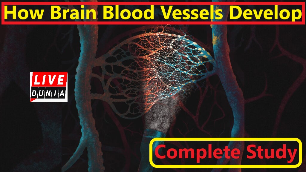
Summary of Blood Supply of the Brain

Brain-specific angiogenic mechanisms activated by terminal cell specialization.
Vertebrates require locally adapted blood vessels. It is often assumed that the advantage of such organotypic vascular specialization has no molecular relationship to organ vascular specialization.
In contrast to this model, here we reveal a molecular mechanism of brain-specific angiogenesis that functions under the control of Wnt7a/b ligands, known signals of blood-brain barrier maturation.
The control mechanism relies on Wnt7a/b-dependent expression of Mmp25, which we found in brain endothelial cells. Mutation of CRISPR-Cas9 in zebrafish reveals that this poorly characterized glycosylphosphatidylinositol-anchored matrix metalloproteinase is selectively required in endothelial tip cells to mediate their initial migration to the brain surface pial basement membrane in the 1990s.
Mechanistically, Mmp25 confers brain invasive potential by cleaving meningeal fibroblast collagen IV-derived α5/6 chains in a small non-collagenous region in the central helix of the heterotrimer.
Genetic interference with pial basement membrane structure abrogates Wnt-β-catenin-dependent organotypic control of brain angiogenesis, resulting in a properly structured but defective blood barrier in the brain vasculature.
We reveal an organ-specific mechanism of angiogenesis, highlighting the mechanistic diversity of tip cells and thereby clarifying that the organ, with spatial constraints on angiogenic tip cells, has its unique physiological needs How they build ships according to account.
The normal blood supply to the brain and spinal cord depends on two groups of branches from the dorsal aorta. The vertebral arteries arise from the subclavian arteries, and the internal carotid arteries are branches of the common carotid arteries. The primary blood supply to the spinal cord comes from the ten cerebral arteries arising from the vertebral arteries and segmental branches of the aorta.
Together, these vertebral arteries form the anterior and posterior vertebral arteries. If one of the vertebral arteries is blocked or damaged (for example, during abdominal surgery), the blood supply to parts of the spinal cord can be disrupted. The type of neurologic damage depends on whether the blood supply to the posterior or anterior artery is disrupted. As expected given the location of ascending and descending nerve pathways in the spinal cord, loss of posterior input usually results in loss of sensory function, whereas loss of anterior input often results in motor deficits.


